Signalment, History, and Physical Exam Findings
 Brennan, a 2 year old, FS, Beagle Hound Mix, ran into a stick that then managed to get stuck in her throat. Her owner was able to pull the stick from her throat, but she then collapsed and was very painful.
Brennan was started on Clavamox and pain medication, but developed significant swelling in her neck over the next few days. When switched to clindamycin, the swelling improved but Brennan continued to be painful. Radiographs were performed and only showed a soft tissue swelling in the ventral cervical region.
She was presented to Dr. Rochelle Anderson of Potomac Valley Veterinary Surgical Specialists four weeks after her injury, was reluctant to move her neck, had a decreased appetite and an occasional cough. On physical examination, Brennan was weight bearing lame on her left forelimb and painful if her neck was manipulated. There was some thickening of the soft tissues in the left ventral cervical area, but no obvious mass, abscess or foreign body was palpable. Brennan Lee’s blood work was normal.
Diagnostics
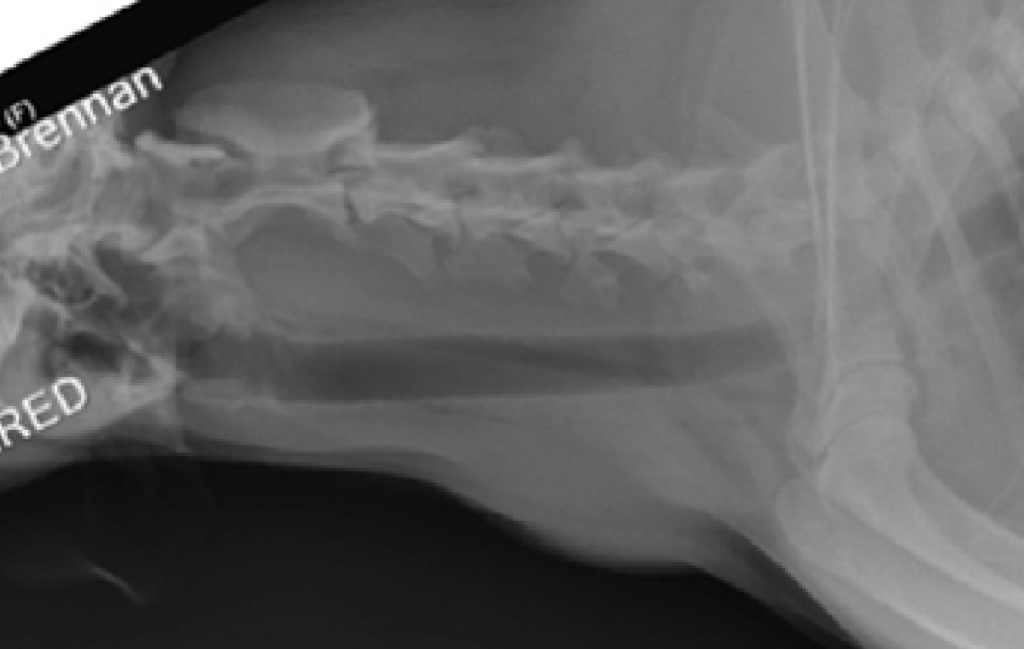
Radiograph showing the soft tissue swelling in the ventral cervical area. A foreign body was not identified.
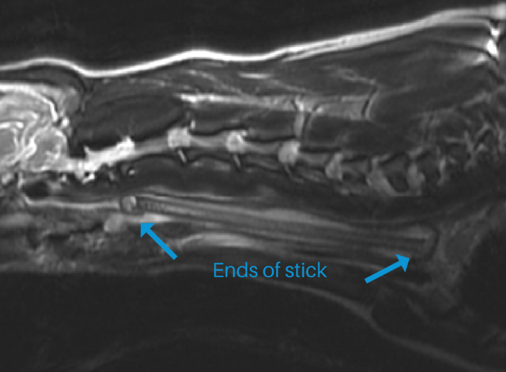
MRI image clearly shows a large foreign body in the cervical region, dorsal and to the left of the esophagus, consistent with a large stick 16cm in length by 8 mm in diameter.
Treatment and Follow-Up
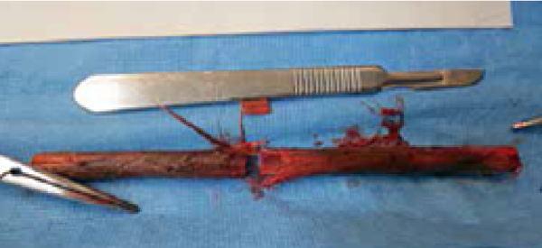
Brennan Lee was taken to surgery immediately following MRI and a stick foreign body was removed.
Brennan has recovered completely and we hear her sister is very happy to have her healthy again!
Conclusion
In retrospect, reviewing the radiograph, a radiolucent linear object can be visualized in a diagonal direction starting below C1/C2 extending to just in front of the shoulder (below the first thoracic vertebrae).
The overwhelming advantage of MRI in this case comes from 2 inherent properties of MRI: True Multiplanar Imaging and Superior Soft Tissue Contrast and Resolution. Because of the superior soft tissue contrast and resolution we are able to easily identify normal from abnormal tissue, not just in this case, but in nearly every case we image. Because of the true multiplanar imaging, we are able to meticulously visualize the foreign body and all of the surrounding tissues. This information is critical as it enables the clinicians to develop a proper surgical plan by knowing what structures are involved prior to attempting removal of the foreign body.
We would like to thank Dr. Rochelle Anderson of Potomac Valley Veterinary Surgical Specialists and Dr. Candice Flynn of Columbia Pike Animal Hospital for referring Brennan Lee to us!!